

What are Oral Tumors in Pets?
ORAL CANCER
Cancer of the oral cavity (mouth) is relatively common in dogs and cats. The annual incidence of oral cancer in dogs is 20 per 100,000 and in cats 11 per 100,000. Although many tumors of the mouth are benign, there are several significant malignant tumors that affect our pets. Dogs are most commonly diagnosed, in decreasing frequency, with malignant melanomas, squamous cell carcinomas, and fibrosarcomas. Cats are most commonly diagnosed with squamous cell carcinomas followed by fibrosarcomas. Because of their location, oral malignant tumors are often detected late in the course of the disease process and have commonly progressed to Stage II at the time of diagnosis. A Stage II tumor is one that is 2-4 cm in diameter with no evidence of further spread.
COMMON SYMPTOMS:
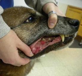
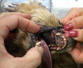
Pets with oral tumors will often have a history of pain while trying to chew or swallow food, food dropping out of the mouth while eating, drooling, or not willing to eat at all. Periodontal disease, bad breath, and tooth loss may also be noted. If lesions are ulcerated, there may be blood-tinged saliva. By the time any abnormality is noticed, the mass is likely to be large enough to be visualized in the oral cavity.
HOW IS THE DIAGNOSIS MADE?
Definitive diagnosis is based on the histopathologic examination of a surgical biopsy of the mass. A fine needle aspirate of the mass can be done initially to get a presumptive diagnosis, but a biopsy is ultimately needed. Staging of the tumor is important before treatment as it governs the treatment plan and helps determine the prognosis. Staging describes if there is bone invasion, local lymph node involvement, or distant metastasis and is evaluated through lymph node aspirates/biopsy and thoracic (chest) radiography.

The size of the tumor can be most accurately determined when a pet is under general anesthesia. At this time a CT (computed tomography) scan can be performed to assess invasiveness into the local tissues. A CBC, chemistry panel, and urinalysis should be evaluated for concurrent disease or organ abnormalities related to metastasis.
Malignant Melanoma:
Malignant melanomas of the oral cavity originate from the mucosa or gingiva. They often have brown or black pigmentation, but can also be non-pigmented. Male dogs with heavily pigmented mucosa such as German Shepherds and Cocker Spaniels may be more predisposed. Malignant melanomas are characterized by rapid growth, local invasiveness, and early metastasis to regional lymph nodes and lungs. Over half of the cases will have bony invasion and lymph node metastasis at the time of presentation and approximately 15% will have detectable lung metastasis. This tumor carries the least favorable prognosis of all of the oral tumors.
Fibrosarcoma:
Fibrosarcomas arise from the gingiva or connective tissue of the hard palate. They often appear firm and smooth with nodules that may become ulcerated and can be seen on the upper jaw between the canine and molar teeth. Dogs with fibrosarcomas are younger (average 7-8 years) than dogs with other oral malignancies (9-11 years). Fibrosarcomas tend to affect large breed dogs. Fibrosarcomas are very locally invasive; therefore recurrence after surgical excision is common. Metastatic behavior is variable.
Non-tonsillar Squamous Cell Carcinoma (SCC):
In cats, these tumors most commonly arise from the gingiva in front of the canine teeth and under the tongue. Other common oral sites include the inside of the cheeks, hard palate, and tongue. They appear as irregular, often ulcerated masses, or raised and inflamed plaques. SCC is characterized by rapid growth, local invasiveness, but tends to be late to metastasize. In dogs bony involvement is particularly common. Regional lymph node metastasis is uncommon (5-10%) Non-tonsillar SCC is the least likely of the oral tumors to metastasize to the lungs (10%), and when found in dogs, have the most favorable prognosis when located in the rostral (front) portion of the oral cavity. In contrast, treatment of cats with SCC is generally palliative with poor success rates.
Tonsillar Squamous Cell Carcinoma:
These tumors are more aggressive than gingival SCC because they are locally invasive AND nearly all have metastasized to the regional lymph nodes at the time of presentation. Potential for metastasis is quite high (>50%). These masses appear as plaque or cauliflower-like lesions often affecting only one tonsil. Tonsillar SCC and SCC occurring in the caudal (back) portion of the oral cavity carry a poor prognosis.
Acanthomatous Ameloblastoma:
These tumors were formerly called acanthomatous epulis. The epulides are a group of benign tumors of dental origin. Acanthomatous ameloblastoma is a member of this group. Although technically “benign” (does not metastasize), it can be aggressive locally by destroying underlying bone. Treatment with surgery and/or radiation therapy is usually curative. Untreated, however, this disease can progress to a point that affected patients can have significant pain in the jaw and are unable to eat.
Treatment of Oral Tumors
Surgery:
Complete local excision with wide surgical margins is recommended. Local recurrence is highly likely with inadequate resection. Large tumors that have invaded the bone may require removal of part of the jawbone in order to obtain “clean” (tumor-free) margins. Early intervention with aggressive surgery can be curative in patients with non-tonsillar SCC and fibrosarcomas. Surgery of small melanomas (e.g. < 2 cm) can result in prolonged survivals.
Radiation therapy:
Some masses are too large to be completely resected, therefore radiation therapy is often recommended in combination with surgery. Some tumors are treated with radiation therapy before surgery in order to attempt to shrink them down to a more operable size. Some patients are treated post-operatively with radiation when the margins of the surgical resection show tumor cells still present.
Chemotherapy:
Chemotherapy may be recommended for patients with more aggressive tumors (e.g. higher grade tumors). In some patients, chemotherapy can provide short-term relief of pain if the patient is considered to have an inoperable tumor or is a poor candidate for radiation therapy.

IMMUNOTHERAPY:
Numerous clinical trials involving the use of melanoma tumor vaccines have been conducted or are underway. Several anti-melanoma vaccines have been evaluated and appear to improve survival for certain patients with this disease. Although apparently safe, efficacy is a concern. It appears that at least 30% of patients with earlier stage disease may benefit from melanoma vaccination.